Non-invasive detection is an evolving field in cancer diagnostics. Optical imaging systems have the potential to achieve this goal due to their non-contact and non-invasive nature, portability, and adaptability. Spectroscopy techniques such as diffuse reflectance, fluorescence, and Raman spectroscopy have gained much attention because they can successfully discriminate cancerous tissues from normal tissues.
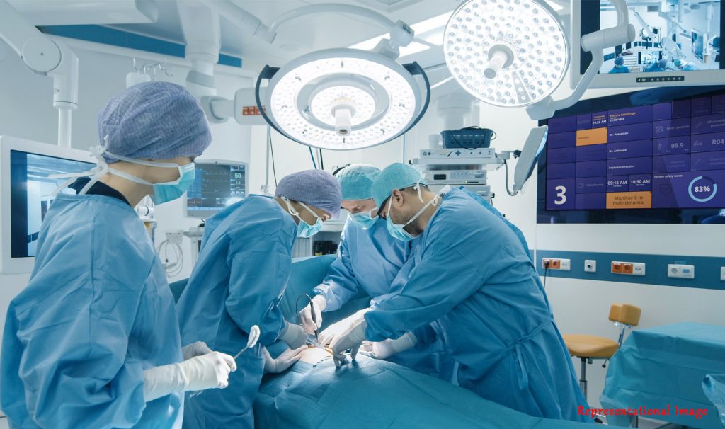
One of the critical challenges in cancer surgery is getting information on tumor margins, which is currently time-consuming during intra-operative procedures. Raman spectroscopy is ideal for classifying different types of cancerous tissues, with a classification accuracy of almost 99 percent. Localizing the tumor’s depth and thickness in operating environments and classification aid in determining acute margins in mastectomy, needle biopsy, and endoscopic applications.

Ms. Subitcha Jayasankar 
Mr. Deepak Bajhaiya 
Prof. Sujatha Narayanan Unni
Conventional Raman spectroscopy has limited penetration depth due to weak signals from the subsurface tissue layers, contributed by the optical interference from surrounding tissues. To enable experimental Raman spectroscopy to counteract these drawbacks, an in-silico study on spatially offset Raman spectroscopy (SORS) is conducted by Ms. Subitcha Jayasankar, Mr. Deepak Bajhaiya, and Prof. Sujatha Narayanan Unni, from the Biophotonics Laboratory, Department of Applied Mechanics, Indian Institute of Technology (IIT) Madras, Chennai, India. Monte Carlo (MC) simulated SORS response coupled with deep learning protocols is employed in this study, promising the use of Raman spectroscopy to locate the tumor’s extent immediately after the surgical excision of the mass.
For the first time, simultaneous soft tissue tumor depth and thickness prediction were carried out with a good root mean square error (RMSE) of 8.4 percent. This study’s depth prediction of the tumor had an RMSE of 4.4 percent, which was better than the 7 to 25 percent RMSE demonstrated by previous reports. This study is expected to help quantify the tumor margins for clinicians to understand the extent of the tumor in the sample before histopathological processing. The methods used in this study could also help the detection of subsurface flat adenoma tumors. Non-invasive tumor development/treatment monitoring is also another benefit of this work.
Dr. Hari M. Varma, Associate Professor from the Biosciences and Bioengineering Department, Indian Institute of Technology (IIT) Bombay, Mumbai, India, acknowledged the importance of this work by giving the following comments: “Identifying the depth at which the cancerous region prevails in a tissue and assessing its size is of utmost importance in cancer diagnosis and treatment. By combining the ability of Raman spectroscopy to detect and classify different cancerous tissues with spatially resolved measurements, together called as Spatially off-set Raman Spectroscopy (SORS), one can achieve deep tissue cancer imaging. However, due to inherent bias of the spatially resolved measurements towards the shallower region of the tissue, accurate depth localization of cancerous region is difficult to achieve resulting in difficulties in surgical intervention. Here, for the first time, authors offer a solution to this problem by incorporating the convolution neural network (CNN) based learning approach to the Monte Carlo based SORS to achieve better depth localization along with improved tumour size assessment. This work, I believe, has a very important clinical application which prompts to have further extensive clinical trials.”
Article by Akshay Anantharaman
Here is the original link to the paper:
https://iopscience.iop.org/article/10.1088/1361-6463/ac8126