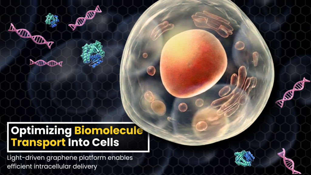
Delivery of biomolecules such as DNA, RNA, and proteins into cells is very important to study cellular processes and to treat diseases through methods like gene therapy and targeted cancer therapies.
The three ways to transfer materials into cells are through viral, chemical, and physical means. In viral methods, the cell’s immune system can recognize the virus’s entry as a threat and act against it. Chemical methods can cause cell damage. Therefore, physical methods are more sought after.
Physical methods for transfecting cells use mechanical, electrical, or optical stimuli to create hydrophilic pores in the plasma membrane through which biomolecules can be delivered into cells before closing the pores. But these methods have their own set of limitations.
For example, mechanical methods have low-throughput (a smaller amount of material able to pass through the cell) requiring highly skilled personnel. Electrical methods cause irreversible cell damage and significantly reduce cell viability (cell viability refers to the proportion of living, and healthy cells in a given sample).
Therefore, photoporation is an alternative to these methods. Photoporation refers to a transfection method where light-matter interaction is used to create temporary nano-sized pores in the cell membrane, and biomolecules are successfully delivered into cells before cell membrane resealing.
Photoporation has several advantages in that it is a high-throughput (larger amount of material able to pass through the cell) intracellular delivery method, does not cause permanent damage to the cells, is a contactless process that reduces the risk of contamination, and yields high cell viability.
Photoporation uses photosensitizing materials such as gold nanoparticles and carbon nanoparticles. This approach offers a high transfection efficiency with high cell viability.
Although the nanophotosensitizer-mediated photoporation method has several advantages, there are problems such as non-uniform delivery, lower delivery efficiency, and lower reproducibility. Furthermore, the uptake of nanoparticles into the cells can result in cytotoxicity (causing destruction of cells), affecting cell viability and biosafety.
Reduced graphene oxide (rGO) is a good alternative to using nanoparticles. Graphene has many good properties, such as superior mechanical, electrical, thermal, and optical characteristics, combined with biocompatibility. This makes them promising in the field of bioengineering and biomedicine.
Reduced graphene oxide is advantageous because of its lower production costs, scalability, reduced degradability, ease of micropattering, and chemical tenability.
Unlike the plasmonic nanoparticles, which show strong absorption at specific wavelengths, the broad and flat spectral characteristics of reduced grapheme oxide range from the visible to the near infrared region, along with its biocompatibility, large specific surface area, and external photothermal properties, making it a good candidate for use as a photosensitizer material for cellular therapy and diagnostics.
In this study, the authors have successfully developed and demonstrated an immobilized, micro-patterned reduced graphene oxide device-based photoporation platform for the fast and efficient intracellular delivery of cargos (transport of a large number of biomolecules) across various cell types, including human mesenchymal stem cells (hMSCs).
By combining the advantages of the photothermal properties of reduced graphene oxide with a spatially defined microarray design, and the precision of near-infrared nanosecond pulse laser-based scanning systems, this platform could achieve controlled cell plasma membrane permeabilization with minimal cell toxicity and exceptional throughput.
The platform used in this study was able to deliver a broad spectrum of biomolecules including PI dye, siRNA, a plasmid for EGFP expression, and enzymes into various cell lines including clinically relevant human mesenchymal stem cells (hMSCs).
The device demonstrates significant potential as a versatile and scalable tool for intracellular delivery, applicable in areas ranging from cell-based therapies, and gene therapy to drug screening and regenerative medicine.
The platform has the ability to transfect a million cells in a few seconds, making it a potential candidate for both in vitro (a process taking place in a glass tube or a dish. Not taking place inside a living organism) and translational medicine applications.
Future studies will focus on integrating this work into in vivo (a process taking place inside a living organism) and 3D tissue culture models, as well as incorporating an automated laser- scanning system with real-time feedback loops for dynamic control of delivery parameters.

The following are the authors of this paper:
- Ms. Donia Dominic from the Department of Engineering Design, Indian Institute of Technology (IIT) Madras, Chennai, India.
- Dr. Rajdeep Ojha from the Department of Physical Medicine and Rehabilitation, Christian Medical College, India.
- Dr. Moeto Nagai from the Department of Mechanical Engineering, Toyohashi University of Technology, Japan.
- Dr. Srabani Kar from the Department of Engineering Design, IIT Madras. Dr. Srabani Kar is also affiliated with the Department of Physics, Indian Institute of Technology (IIT) Hyderabad, India.
- Dr. Tuhin Subhra Santra from the Department of Engineering Design, IIT Madras. Dr. Tuhin Subhra Santra is also affiliated with the School of Interdisciplinary Studies, IIT Madras, Chennai, India.
Prof. Hwan You Chang, Emeritus Professor at the Institute of Molecular Medicine, National Tsing Hua University, Hsin Chu, Taiwan, pointed out the importance of the work done by the authors with the following comments: “This article addresses a critical bottleneck in cell biology and therapeutics: the need for a delivery method that is simultaneously high-throughput, efficient, and compatible with a wide range of cargo types and cell lines. The authors have devised an elegant platform that achieves these objectives while offering the additional advantage of avoiding undesired interference from the photosensitizers. I anticipate that commercialization of this system would greatly benefit the broader cell biology community.”
Article by Akshay Anantharaman
Click here for the original link to the paper