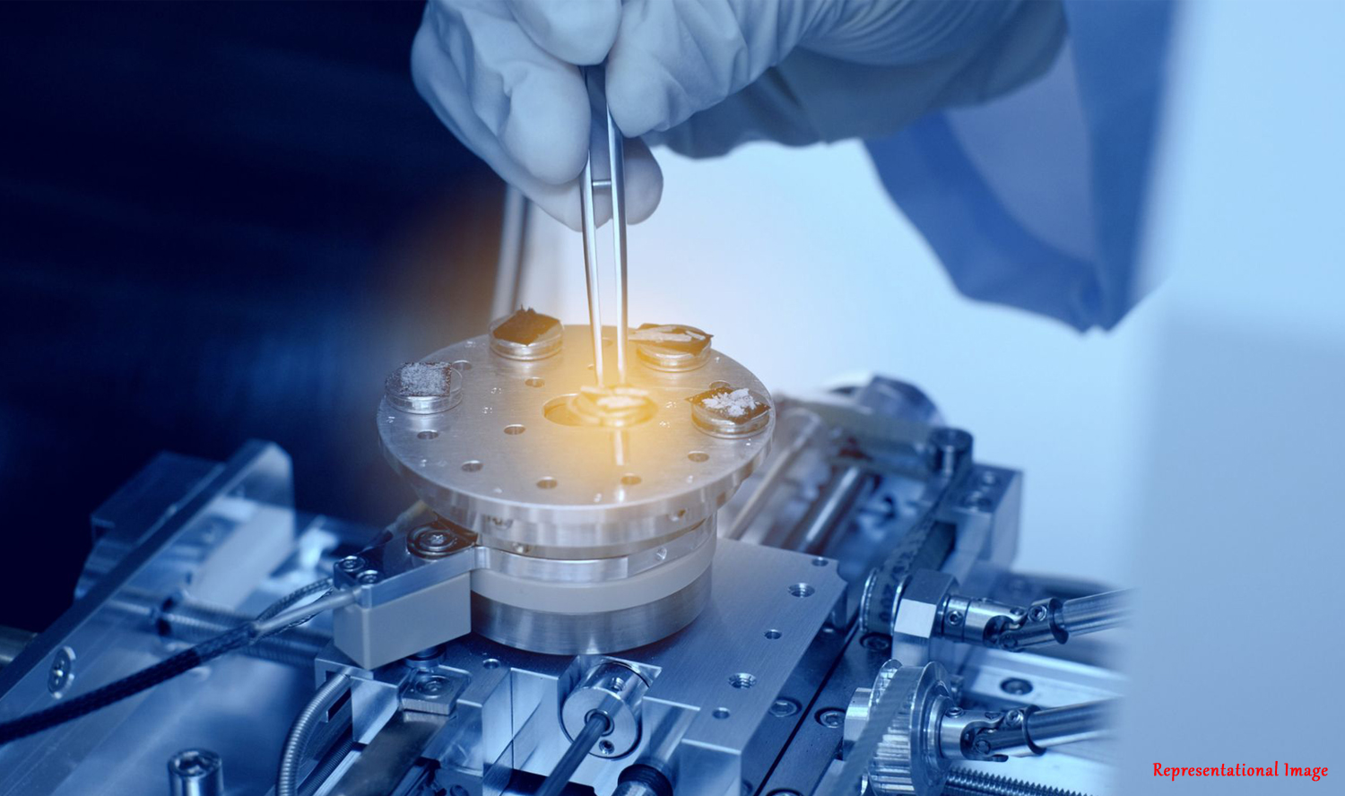
Can we detect cancer and other malignant cells using a handheld device like the ones being used for pregnancy tests and diabetes? Recent work from Prof. Pushpavanam’s lab at IIT Madras in collaboration with researchers at Clemson University, USA boosts confidence that in the near future ‘we can’. Known as Lab-on-Chip devices, these consist of a channel of characteristic dimensions of a few hundred micrometers through which extremely small volumes of fluid flow. These miniature devices, conceptually akin to blood-glucose monitors, can be envisioned to detect malignant cells in peripheral blood.
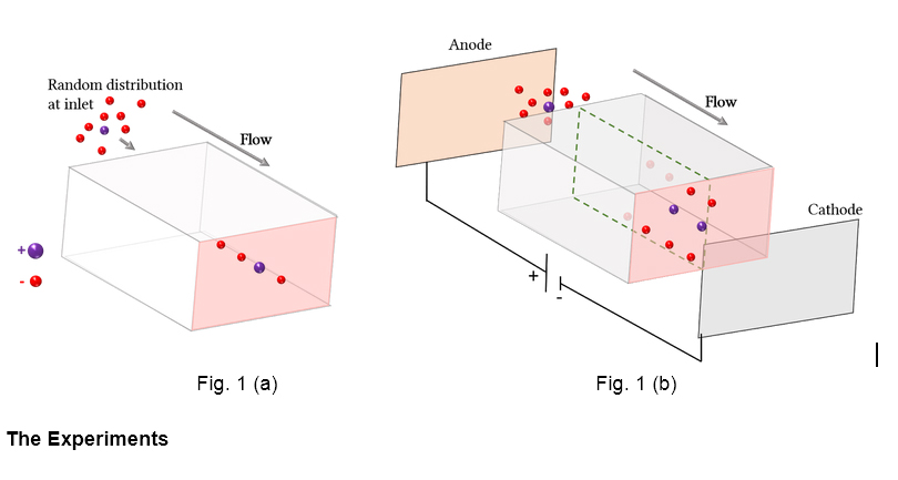
It was observed in the 1960s that millimeter sized particles in a dilute suspension arrange themselves when passed through channels. This also applies to micro-particles and other minute cells. This means that cells and micro-particles within the flow can potentially separate themselves out without employing any membrane. But that separation depends upon many parameters of the cells and channel, and may not be significant for any practical applications {Fig. 1(a)}. What if, by any means, we can make those separations more distinct, which might be helpful for early disease detection? This is what the IIT Madras and Clemson University teams predicted theoretically and verified experimentally.
Biological fluids, being non-Newtonian, do not follow Newton’s law of viscosity. Hence they exhibit elastic and viscous behavior. In this context, applying weak electric fields parallel to the flow of a non-Newtonian fluid suspended with micron sized particles, results in them either focusing at the centerline of the microchannel or near the walls, depending upon the polarity of the electric field {Fig. 1 (b)}.Electric fields thus allow active control over the particle focusing position.
The Experiments
Liquids in our body are mostly non-Newtonian. To mimic them, a dilute solution of extended molecular chains (polymers) was prepared, and micrometer sized polystyrene particles were suspended representing various cells. Similar to our childhood experiment of running a comb in dry hair which imparts an electric charge on the comb, polystyrene particles attain negative charges in the solution. Choosing polystyrene particles was motivated by cancer cells; they maintain a strong negative charge on their surface which facilitates its multiplication and growth. These electrostatic charges are the key to their detection and more focused separation.
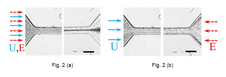
The researchers at Clemson University found that, applying an external electric field in the direction of the fluid flow resulted in focusing the polystyrene particles to the centerline of the channel {Fig. 2 (a)}. Whereas, upon reversing the direction of the electric field, polystyrene particles were found to focus near the walls {Fig. 2 (b)}. “Owing to the non-linear nature of viscoelastic flow, a weak focusing of particles was there before as well; the electric field just accelerated it,” said Akash Choudhary, doctoral scholar in the Department of Chemical Engineering, IIT Madras, who led the study. “Furthermore, the electric field provides an external control over the particle migration and enables charge-selective focusing” he added.
Generally, a charged particle migrates along the line of an electric field. But here, we see them migrating in the transverse direction as well. Why is that so?
The Theoretical Model
To understand this, researchers at IIT Madras created a theoretical model of their system. They started with fundamental fluid dynamic and electrostatic equations for a Newtonian fluid and approached the non-Newtonian behaviour step by step, by introducing the elastic nature of the flow as a small perturbation. Like the negatively charged comb attracts paper bits by inducing upon them a positive charge, the negatively charged polystyrene spheres get surrounded by a thin blanket of positive charges in an aqueous medium. Upon the application of an electric field, this blanket slips instantaneously, resulting into an electrokinetic motion of polystyrene particles. This motion generates disturbances, i.e., ripples in the fluid medium.
Furthermore, as the polystyrene particles are rigid solids, they modify the flow, causing further disturbances. Interaction of all these ripples, i.e. the electrokinetic disturbances and the fluid disturbances due to the rigidity of the polystyrene particles, results in the transverse motion of the particles which manifests as focusing. How much focusing did this method achieve? Upon solving analytically the governing equations, researchers found that an order of magnitude enhancement in focusing is obtained. Both the theoretical results and experimental analysis were found to be in good agreement. Now, when can we detect cancer cells at the earliest?
Challenges
In the case of most carcinomas, the concentration of circulating cancer cells in the bloodstream is very low; just one cell present among ten crores of red blood cells. This renders the separation difficult. Findings from the current research suggest that negative surface charge of rare cancer cells can be used to enhance detection via an application of weak electric fields parallel to the flow. But, “making such a lab-on-chip device for cancer cells detection is a long-term goal – the prime motivation for fathoming major challenges” Akash said, “A starting building block, I would describe this study as” he added.
Collaborators

Akash Choudhary 
Di Li 
T. Renganathan 
Xiangchun Xuan 
S Pushpavanam
Article by Mywish Anand
Here is the link to the research article:
https://www.cambridge.org/core/journals/journal-of-fluid-mechanics/article/electrokinetically-enhanced-crossstream-particle-migration-in-viscoelastic-flows/E28632EB7D8952C0003BDCAEB30716BE